

Home > Company News
Nasal endoscopy provides a platform for precise visualization and treatment, but the actual hemostatic method depends on the site, vessel size, bleeding rate, mucosal condition, previous treatment, and the patient's systemic risk. Clinicians generally begin with the least traumatic focused option that is likely to work and escalate when local measures do not provide stable control.
Targeted micro-packing places an absorbable hemostatic product or a small packing material directly over a defined bleeding area under endoscopic vision. It can support hemostasis in focal oozing, at a site that is not suitable for immediate cautery, or when clot formation needs local assistance. Compared with packing the entire nasal cavity, focused placement often has less effect on airflow, although product choice, pressure, and removal timing remain clinical decisions.
When the source has not been identified, simply adding more packing may conceal posterior bleeding and increase pain, mucosal pressure injury, or infection risk. Finding the location is more important than filling more space.
Chemical cautery is commonly used for a small superficial anterior vessel, while bipolar electrocautery can provide focused control of a clearly visible vascular stump. Both require a clean view and should not be repeated broadly when the source is uncertain. Septal mucosa is thin; treatment that is too deep, too extensive, or applied to matching areas on both sides may increase crusting, adhesions, and perforation risk.
Heavy active bleeding can prevent accurate delivery of energy. Suction, pressure, or clinician-directed topical medication may first be needed to improve the field before limited coagulation is performed.
These instruments use different forms of energy to coagulate tissue and seal a bleeding vessel. They may be selected for particular deep or recurrent sources, but more power and longer application are not inherently better. The clinician adjusts the device according to the generator, probe, tissue response, and anatomic site, usually using brief and confined applications.
Extra care is required near septal cartilage, alar skin, the orbit, and the skull base. A fixed power or distance cannot be treated as a universal standard apart from the specific device instructions and clinical context.
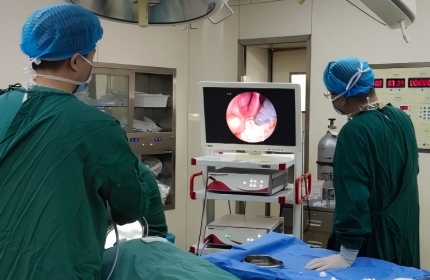
For recurrent high-volume posterior epistaxis, failure of appropriate local treatment and packing, or a suspected arterial source, the ENT team may consider surgical control such as endoscopic sphenopalatine artery ligation. Endovascular embolization is reserved for selected cases after imaging and multidisciplinary assessment, including some refractory arterial bleeds or vascular lesions. Both options have specific indications and risks and are not routine responses to an uncomplicated nosebleed.
• Whether the source can be seen clearly and whether active bleeding is ongoing.
• Whether bleeding arises from superficial capillaries, a small arterial stump, or a larger posterior vessel.
• Use of anticoagulants and the presence of a blood disorder, hypertension, or another anesthesia risk.
• Failure of previous packing, cautery, or surgery and the current condition of the nasal mucosa.
• Available equipment and the clinician's training and experience with the selected technique.

+86-13167065777
Building 5A, No. 9628, Hunan Highway, Pudong New Area, Shanghai, China
 Links:
Shanghai Shiyin Photoelectric Instrument Co.,Ltd. All Right Reserved